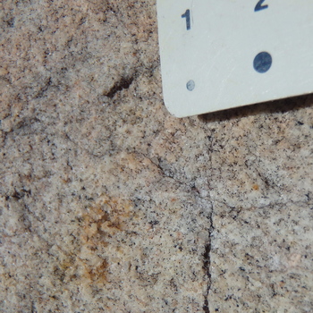
1.0 Aplitic granite. Carson-Iceberg Wilderness, California. [Scale in upper right shows 1 and 2 mm diameter dots.] Gray quartz is especially visible near intersecting cracks below the 1 mm dot. Alkali (K-rich) feldspar is pink. |
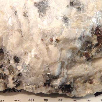
2.0 Coarse-grained garnet-muscovite granite. McKinney Mine, Mitchell County, North Carolina. Gray quartz, white alkali (K-rich) feldspar, black to silvery-clear appearing muscovite mica, and red garnets. [Scale in mm and cm, with 10 mm intervals.] |
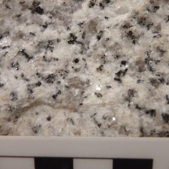
2.1 Biotite granite. Near Woodleaf, North Carolina. Gray quartz, white alkali and plagioclase feldspars, and black biotite. [The scale is the standard GSA scale with cm divisions.] |
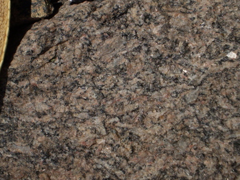
3.0 Sillimanite-biotite granite. Allenspark, Colorado. Gray quartz, pink alkali (K-rich) feldspar, white plagioclase feldspar, and black biotite. Sillimanite forms tiny (invisible) needles near and within biotite grains. [The part of the hat rim showing in the upper left, providing scale, is 8 cm long.] |
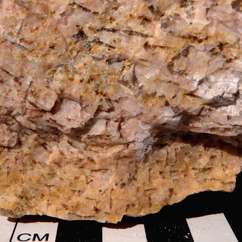
4.1 Graphic pegmatitic granite. Custer, South Dakota. Glassy, reddish gray quartz occurs as hieroglyphic-like markings within pink-orange alkali feldspar. The feldspar is a single large grain of more than 10 cm. All quartz is likely the interconnected parts of a single, skeletal crystal. |
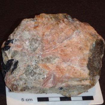
4.2 Pegmatitic tourmaline-muscovite granite. Custer, South Dakota. Gray, glassy quartz, black to silvery-white muscovite mica, and black tourmaline decorate a large, orange-pink crystal of alkali (K-rich) feldspar. |
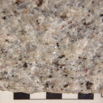
5.0 Biotite quartz monzonite. Salisbury, North Carolina. Gray quartz, pink alkali feldspar, white to pink plagioclase feldspar, black biotite and magnetite, and red to orange hematite comprise the rock. [Scale at right is standard GSA scale with cm divisions.] |
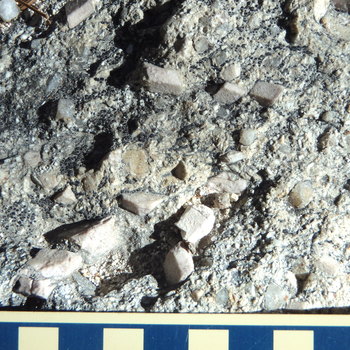
5.1 Weathered, porphyritic quartz monzonite. Castle Crags, California. Gray glassy quartz forms square-appearing euhedral crystals that project above the surface along with the more dominant, euhedral, pink alkali feldspar crystals. Plagioclase feldspar occurs as smaller white crystals. Black bumpy spots are lichens growing on the weathered surface. [Scale is standard GSA scale with cm divisions.] |
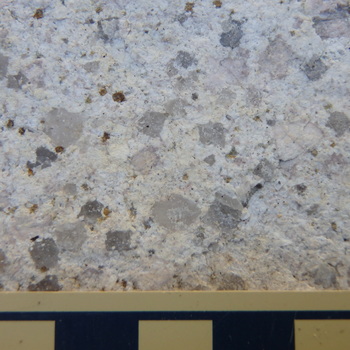
6.0 Porphyritic leuco-granodiorite. Castle Crags, California. Gray glassy quartz forms square-appearing euhedral crystals, euhedral pink crystals are alkali feldspar, and white crystals are plagioclase feldspar. A weathered mafic phase appears as goethite or limonite fillings in small depressions. [Scale is standard GSA scale with cm divisions.] |
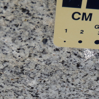
6.1 Hornblende-biotite granodiorite. Sierra Nevada Batholith, US Highway 50 east of Kyburz, California. The rock consists of gray glassy quartz; small, white plagioclase feldspar grains; large, white, poikilitic alkali feldspar crystals; equant black biotite grains; and rectangular to diamond-shaped, black hornblende crystals. Just below the scale, an amoeboid poikilitic grain of alkali feldspar reflects light from its surface. |

6.2 Biotite-hornblende granodiorite. Slightly weathered. Carson-Iceberg Wilderness near Mosquito Lake, California. Minerals the same as in 6.1, but alkali feldspars are smaller and a clearly zoned, large, plagioclase feldspar grain is present in the lower left center. Equant biotite and diamond-shaped to rectangular hornblendes are subequal. Orange stain on left is limonite or goethite. [Scale is standard GSA scale with cm divisions.] |
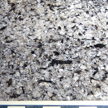
6.3 Biotite-hornblende granodiorite. Carson-Iceberg Wilderness near Mosquito Lake, California. Minerals the same as in 6.2, but hornblende is more abundant and a few occur as larger crystals, notably the rectangular one in the lower center. Plagioclase tends to be generally more subhedral. [Scale is standard GSA scale with cm divisions.] |
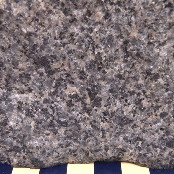
6.4 Hornblende-biotite granodiorite. St. Cloud, Minnesota. Pink alkali feldspar is mixed with gray quartz, plagioclase feldspar, black biotite, and hornblende. [Scale is standard GSA scale with cm divisions.] |
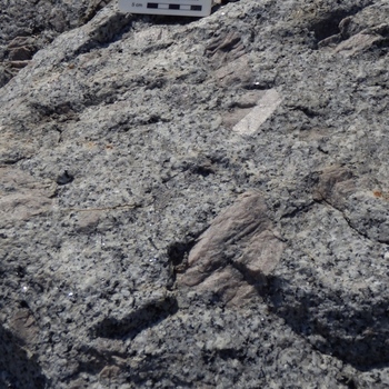
6.5 Porphyritic hornblende granodiorite, Yosemite National Park, California. Note the cluster of large Carlsbad-twinned pink alkali feldspars in a matrix of white plagioclase and gray quartz, with scattered grains of rectangular black hornblende. Other minerals such as biotite and titanite are not readily discernible. |
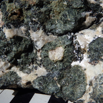
6.6 Orbicular hornblende-augite granodiorite. Davie County, North Carolina. Orbs consist of quartz-epidote cores rimmed by augite (lighter, duller green) and hornblende (dark green to black) in a matrix of gray to white, glassy quartz and white to orange-white alkali feldspar with a few small white grains of plagioclase feldspar. Titanite and orange-iron oxide minerals occur in some orb centers. Titanite occurs as adamantine, yellow to brown crystals (e.g., upper left and mid-right). Nearly invisible are yellow to light red, needle-like grains of rutile within feldspars. [Scale is standard GSA scale with cm divisions.] Commonly, in outcrop, plagioclase dominates over alkali feldspar, so that such rocks are quartz diorites. |
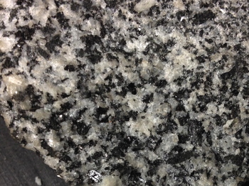
7.0 Hornblende-biotite quartz diorite. Locality unknown. |
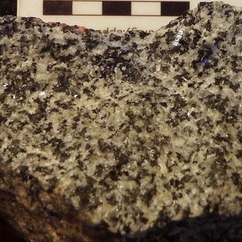
7.1 Hornblende-biotite quartz diorite (tonalite). Alpine, San Diego Co., CA. White, irregular to rectangular plagioclase with gray glassy quartz surround yellow-green epidote, greenish-black rectangular hornblende, and small to large poikilitic brownish-black biotite grains in this rock. Note the large amoeba-like biotite in the upper center containing enclosed grains of quartz and plagioclase. Another poikilitic biotite present at the upper center far left shows reflections from cleavage. In the upper right and elsewhere, rectangular plagioclase grains, including a zoned grain at the right center, show bright reflections from cleavage. |
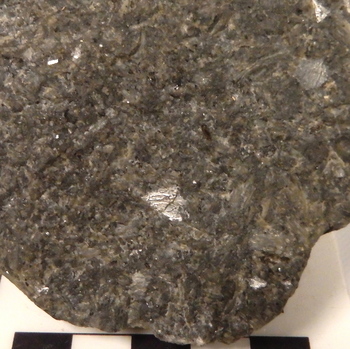
8.0 Syenite. Locality unknown. White to greenish-brown and grayish brown alkali feldspars are associated with minor black biotite mica. Sample courtesy of Sonoma State University Geology Department. [Standard cm scale provided.] |
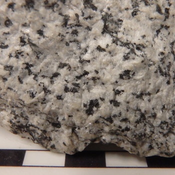
8.1 Quartz-bearing hornblende syenite. Locality unknown. Gray, glassy quartz is scattered between white, gray, and pinkish gray alkali feldspar grains and a few similarly colored plagioclase grains. The dominant black minerals are hornblende, which occurs as irregular to rectangular grains, and biotite appearing as more equant, irregular grains. In addition, some magnetite or ilmenite is visible as tiny, black metallic grains. [Standard cm scale provided.] |
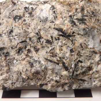
8.2 Hornblende syenite. West Texas. Alkali feldspars are white to gray to light-orange, but are stained darker orange in some places. Gray grains include some plagioclase. Hornblende forms radiating clusters and individual rectangular to wedge-shaped grains. Tiny magnetite or ilmenite grains make black spots surrounded by oxidation rings. Pink, red, orange, and yellow mineral coatings of hematite and limonite (?) surround hornblendes and other iron-bearing grains. [Standard cm scale provided.] |
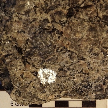
8.3 Aegirine syenite. Concord, North Carolina. Brown, orange, yellow, and greenish white subhedral to anhedral alkali feldspars, with small greenish black aegirine pyroxenes, black biotite, and minor silvery black magnetite comprise the rock. |
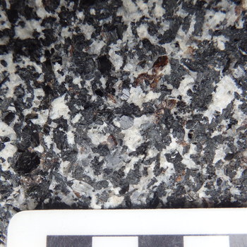
9.0 Titanite-bearing, biotite-hornblende diorite. Yosemite Valley, Yosemite National Park, float block. Adamantine, honey-brown wedges of titanite, and subhedral to euhedral books of biotite are scattered amidst abundant black hornblende and white plagioclase grains. |
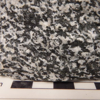
9.1 Hornblende diorite. Locality unknown, but rock is similar to diorite of Woodleaf, North Carolina. The dominant minerals of this rock are black to dark green hornblende and glassy gray to white plagioclase feldspar. Grains of magnetite, ilmenite, or both occur as tiny, scattered grains. [Standard cm scale provided.] |
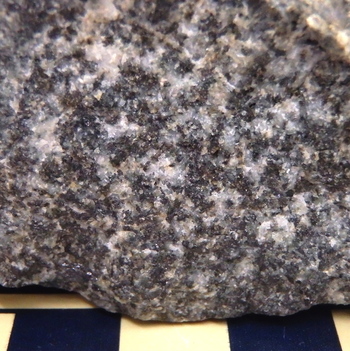
9.2 Hornblende diorite. Salem, Massachusetts. White to dark gray plagioclase feldspar and brownish-black to black hornblende, both with generally irregular grain boundaries, dominate the rock. Sample courtesy of Sonoma State University Geology Department. [Standard cm scale provided.] |
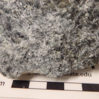
10.0 Augite-hornblende-bearing anorthosite. Locality unknown. Medium- to coarse-grained, white to greenish gray and gray plagioclase and dark green to black augite dominate the rock. Minute iron-bearing oxide grains are minor accessory minerals. Sample from Sonoma State University Geology Collection. [Standard cm scale provided.] |
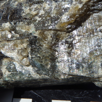
10.1 Phlogopite orthopyroxene anorthosite. Nain, Labrador. The rock is phaneritic to pegmatitic and composed predominantly of labradorite plagioclase feldspar showing labradorescence (a play of colors in the feldspar). Accessory phlogopite and orthopyroxene, both dark brown to black, fill inter-grain areas between large plagioclase grains of white, brown, greenish-brown, and brownish black color. [Standard cm scale provided.] |

11.0 Gabbro. Middletown, California. This rock is a medium-grained gabbro consisting of white to green and dark greenish gray plagioclase feldspar with interspersed dark green to black augite and trace amounts of tiny pyrite grains. [Scale is standard GSA scale with cm divisions.] |
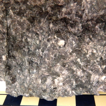
11.1 Gabbro. Bishops Castle, Shropshire, England. Brownish gray plagioclase surrounds dark green augite and minor Fe-bearing oxide minerals. Sample courtesy of Sonoma State University Geology Department. [Scale is standard GSA scale with cm divisions.] |
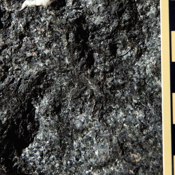
11.2 Hornblende gabbro. Del Puerto Canyon, NE Diablo Range, California. Gray, greenish-gray, and white plagioclase is intergrown with abundant black hornblende. [Scale is standard GSA scale with cm divisions.] |
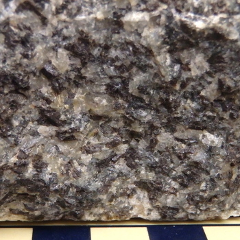
13.0 Norite (hypersthene gabbro). Stillwater Complex, Montana. White, pinkish gray to gray plagioclase feldspar and brownish black orthopyroxene comprise the bulk of this rock and all of the identifiable minerals visible here. [Scale is standard GSA scale with cm divisions.] |
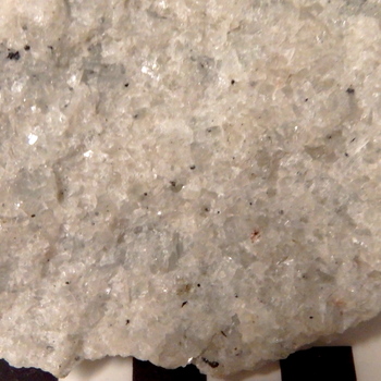
14.0 Nepheline syenite. Locality unknown. Gray nepheline, pinkish white alkali feldspar, and minor iron-bearing oxide minerals, locally weathered to red hematite, comprise the rock. [Scale shows cm divisions.] |
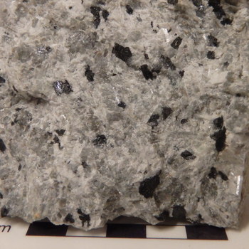
14.1 Sodalite-hastingsite nepheline syenite. Red Hill Complex, New Hampshire. Gray nepheline and greenish-gray sodalite are associated with abundant white to medium gray alkali feldspar, locally showing pronounced cleavage. The black, rectangular grains are the alkali amphibole, hastingsite, and a few metallic grains of magnetite occur as minor accessory minerals. [Scale shows cm divisions.] |
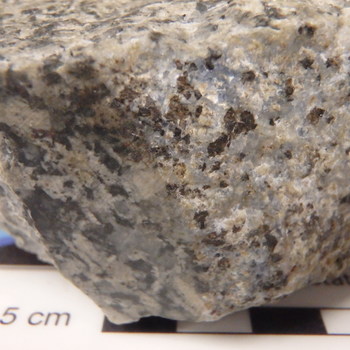
14.2 Sodalite-veined, nepheline syenite. Locality unknown. Blue sodalite, white alkali feldspars, and red-brown weathered alkali amphiboles form a vein now present on the edge of the sample. On the top side of the sample, waxy nepheline and white alkali feldspar dominate the rock. [Scale shows cm divisions.] |
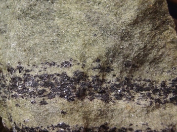
15.0 Chromitite layer in (meta)dunite. Day Book Mine, North Carolina. Chromite grains are dominantly <1 – 2 mm and are locally rimmed by the purplish silver chromium chlorite, kammererite, in the chromitite. In the (meta)dunite and chromitite, the olivine of the (meta)dunite is granular and green of various shades and is accompanied by nothing other than chromite and kammererite. The latter mineral and elongation of chromite pods provides the only evidence of metamorphism. |
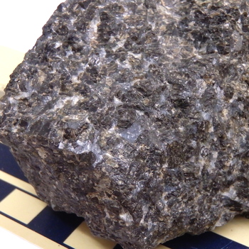
16.0 Harzburgite. Stillwater Complex, Nye, Montana. Minor bluish gray to greenish black plagioclase accompanies minor green olivine and abundant brownish black orthopyroxene in the rock. Sample courtesy of Sonoma State University Geology Department. [Scale is standard GSA scale with cm divisions.] |
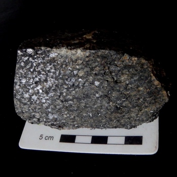
16.1 Harzburgite. Stillwater Complex, Montana. A single large orthopyroxene grain (the oikocryst) encloses irregular chadacrysts of brownish to greenish-black olivine to form an ophitic texture. The sample was photographed with reflection on the left end coming from the cleavage surfaces of the single large orthopyroxene, but the nonreflective surface of the rock at the right shows the normal dark, greenish-black appearance of this harzburgite. |
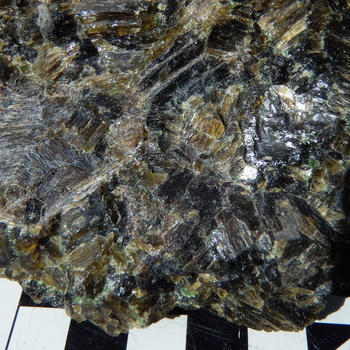
17.0 Websterite. Webster, North Carolina. Bright green chromian diopside accompanies black, deep brown, brown, and greenish brown orthopyroxene. [Scale shows cm divisions.] |
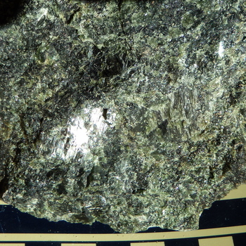
18.0 Clinopyroxenite. Del Puerto Canyon, California. This sample is composed almost entirely of light to dark green augite. Traces of olivine are present. [Scale is standard GSA scale with cm divisions.] |
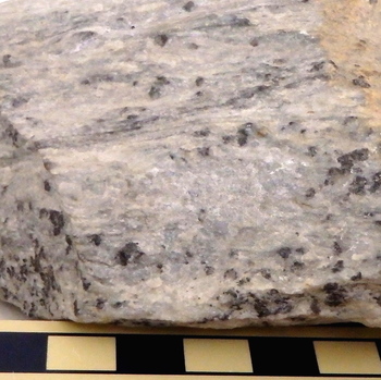
19.0 Carbonatite. Calcite (?) and minor alkali amphiboles constitute this rock. Other possible carbonate minerals are ankerite and dolomite. This sample courtesy of Sonoma State University Geology Department. [Scale is standard GSA scale with cm divisions.] |
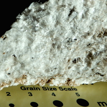
20.0 Pumiceous porhyritic rhyolite. Chimaltenango region, Guatemala. Clear glass fibers, gray quartz phenocrysts, and minor black biotite phenocrysts constitute this vesicular rock. |
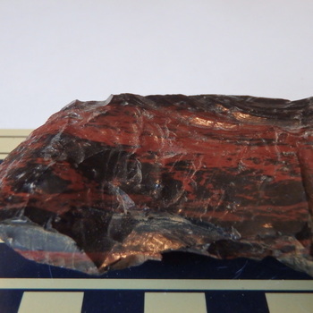
20.1 Rhyolite obsidian. Clear Lake area, California. [Scale is standard GSA scale with cm divisions.] |
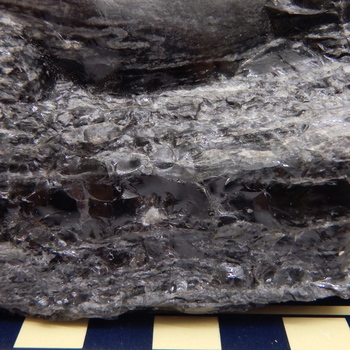
20.2 Black rhyolite obsidian. Locality unknown. Locally pumiceous white glass is surrounded by the dominant black volcanic glass. One glassy round quartz (?) phenocryst is present in the upper left and one granular alkali feldspar (?) clot forms a white patch in the lower middle of the sample. Note the folded layers. Sample courtesy of Sonoma State University Geology Department. [Scale is standard GSA scale with cm divisions.] |
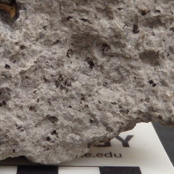
21.0 Vesicular porphyritic rhyolite. Chafee County, Colorado. A granular aphanitic matrix containing scattered vesicles encloses black biotite and hornblende phenocrysts, as well as gray quartz and white alkali (K-rich) feldspar phenocrysts. Sample courtesy of Sonoma State University Geology Department. [Scale shows cm divisions.] |
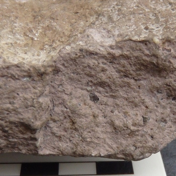
21.1 Porphyritic rhyolite. Churchill County, Nevada. Pink, granular aphanitic matrix encloses gray, glassy quartz phenocrysts and a few white alkali feldspar (sanidine -?) phenocrysts. Sample courtesy of Sonoma State University Geology Department. [Scale shows cm divisions.] |
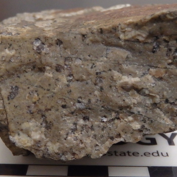
21.2 Porphyritic rhyolite. Salem, Massachusetts. Phenocrysts of gray to red, hematite-stained quartz and white alkali feldspar and small phenocrysts of black to reddish-black weathered ferromagnesian minerals occur in the red-stained crystalline matrix of this rhyolite. Sample in Sonoma State University Geology Department Collections. [Scale shows cm divisions.] |
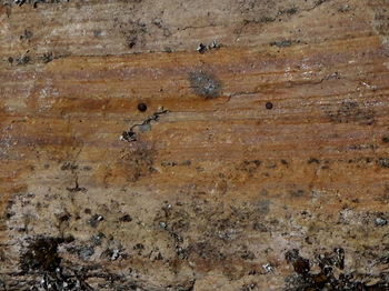
21.3 Flow-banded, porphyritic rhyolite. Annadel State Park, California. Thin layers are millimeter scale. Small rectangular to equant phenocrysts of white alkali feldspar and equant gray quartz, plus small black phenocrysts of ferromagnesian minerals, occur in a grainy flow-laminated, aphanitic matrix. |
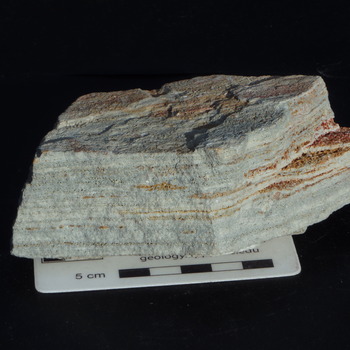
21.3.1 Slightly porphyritic, flow-banded alkali rhyolite. Nuns Canyon, Kenwood, California. Goethite (?) coats orange-weathered, vesicular, flow-band surfaces on which minor, tiny aegirine crystals are present as black grains. A few sanidine crystals form small phenocrysts, one of which, in the upper center of the sample, reflects light from a cleavage surface. |
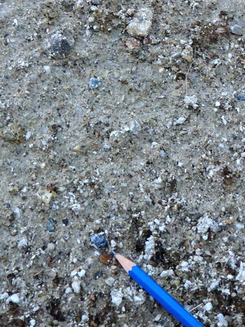
21.4 Lapilli tuff rhyolite. Poorly lithified. Annadel State Park, California. Gray matrix of aphanitic to very fine-grained volcanic ash encloses lapilli of various volcanic rock types and quartz. [Sharpened end of pencil approximately 1.5 cm for scale.] |
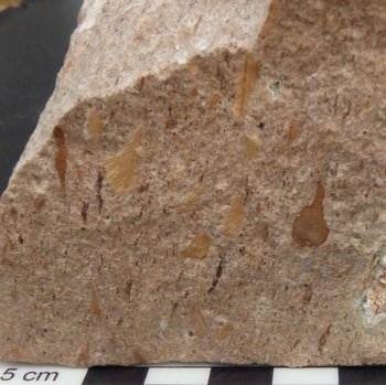
21.5 Welded lithic lapilli tuff rhyolite. Near Shoshone, California. Generally brown volcanic lithic clasts (lapilli) and some crystals of quartz are embedded in an annealed ash matrix of volcanic glass, crystals, and lithic fragments. |
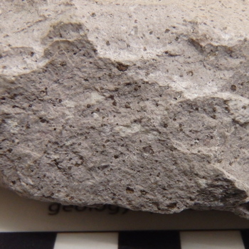
22.0 Quartz latite. Locality unknown. Weathered, red-brown biotite phenocrysts and a few clear to white sanidine (alkali feldspar) phenocrysts are surrounded by a pinkish-purple, aphanitic, crystalline groundmass. [Scale shows cm divisions.] |
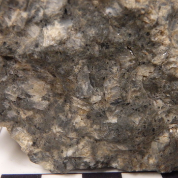
23.0 Porphyritic trachyte. Devils Tower, Wyoming. Large white to clear phenocrysts of alkali feldspar (anorthoclase) and small phenocrysts of clinopyroxene (augite and aegirine) crowd the aphanitic matrix. [Scale shows cm divisions.] |
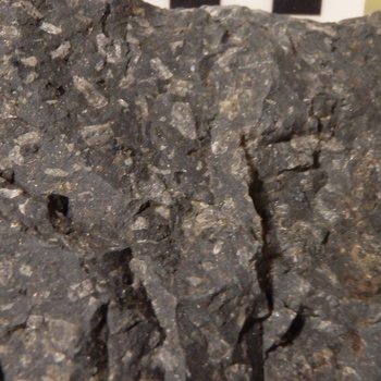
24.0 Porphyritic hornblende latite. Jamestown, California. White phenocrysts of feldspars and brown-weathered hornblende occur in the dark gray aphanitic matrix. [Scale is standard GSA scale with mm diameter dots of 1 mm to 5 mm diameter.] |
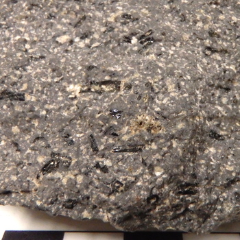
25.0 Porphyritic hornblende andesite. Sierra Nevada, California. Black, rectangular hornblende phenocrysts, plus cream to white plagioclase phenocrysts, are scattered within the gray aphanitic groundmass. [Scale shows cm divisions.] |
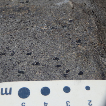
25.1 Porphyritic hornblende andesite. Mosquito Lake, California. Tiny to 3 mm aligned and elongate black phenocrysts of hornblende and a few glassy white to cream-colored, commonly rectangular, phenocrysts of plagioclase characterize the phyric grains of this rock, which has an aphanitic, dark gray groundmass. [Scale is standard GSA scale with mm diameter dots of 2 mm to 5 mm diameter.] |
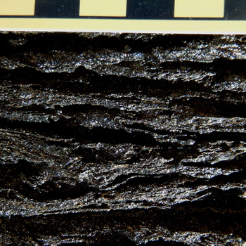
26.0 Ropy glassy basalt. Hawaii. Black, locally folded and layered basaltic glass of pahoehoe character distinguishes this sample. [Scale is standard GSA scale with cm divisions.] |
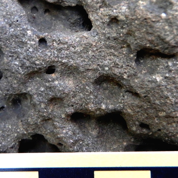
26.1 Vesicular porphyritic basalt. Hat Creek-Mt. Lassen area, California. The large vesicles and rectangular white plagioclase crystals are the most obvious features of this basalt, but some tiny augite (?) phenocrysts also are present in the gray, aphanitic groundmass. [Scale shows cm divisions.] |
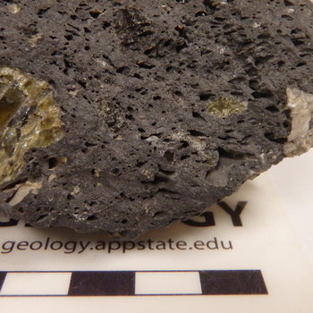
27.0 Vesicular, porphyritic olivine basalt. Locality is near Elko, Nevada. Large glassy green to black phenocrysts of olivine and a few white phenocrysts of plagioclase (one, on the far right, shows cleavage) scattered in the gray aphanitic matrix and among the numerous vesicles, characterize the rock. Sample courtesy of David Bero. [Scale shows cm divisions.] |

28.0 Kimberlite. Kimberley, South Africa. The rock is basically a breccia of angular fragments of various rock types in a serpentine-calcite matrix containing a variety of other (not visible) minerals. Vesicles are abundant. [Scale is standard GSA scale with cm divisions.] |
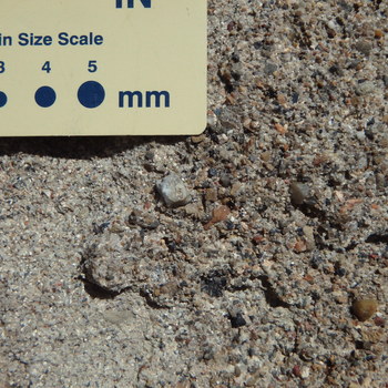
29.0 Polymict granule conglomerate. Dinosaur National Monument, Utah. Multi-colored sand (< 2 mm in diameter) matrix and granules (2–4 mm in diameter) of various rock types, including chert, and minerals, mostly quartz, comprise this conglomerate. Some cement, largely invisible, is present. [Scale is standard GSA scale with mm diameter dots of 3 mm to 5 mm diameter.] |

29.1 Oligomict quartz-pebble conglomerate. Little Mountain, Broadford Quadrangle, Virginia. Well-rounded granules and pebbles (2–10 mm in long dimension) of quartz in quartz-sand matrix constitute this conglomerate. [Scale is standard GSA scale with mm diameter dots of 3 mm to 5 mm diameter.] |
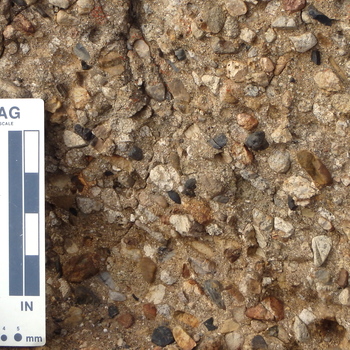
29.2 Polymict pebble conglomerate. Stillwater Cove Regional Park, Sonoma County, California. This conglomerate is characterized by abundant igneous rock clasts (such as the light granitoid clast in the upper left between the two black clasts and the fairly round, cream colored granitoic clast in the center of the field of view) and quartz, but the conglomerate also contains limestone, volcanic rock, and metamorphic rock clasts. The matrix is muddy sand. [The scale is the standard GSA scale with mm and inch divisions.] |
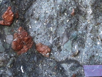
29.3 Polymict pebble to cobble conglomerate. Russian Gulch, Sonoma Coast State Park, California. This conglomerate is distinctive in its inclusion of clasts of red radiolarian chert, green chert, and green mafic volcanic rock clasts, but it includes a variety of sandstone and other rock clasts, as well. Clast shapes are well rounded to subangular. [The scale shown is about 6 cm in length.] |
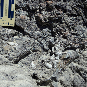
29.4 Fossiliferous pebble-cobble conglomerate (note dinosaur bone). Dinosaur National Monument, Utah. Red, green, gray, and black chert plus other rock types and dinosaur bone fragments constitute the clasts in this sandstone-matrix conglomerate, which has abundant calcite cement. [The scale is the standard GSA scale with mm and inch divisions.] |
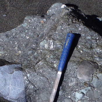
29.5 Boulder-cobble conglomerate. Sonoma Coast State Park, California. This conglomerate shows a wide range of clast sizes from granules to boulders (such as the > 30 cm blue glaucophane schist boulder at the left). Clasts include a wide range of rock types including green mafic volcanic rocks, chert, and gray sandstones. [The part of the hammer handle showing is approximately 33 cm long, for scale.] |
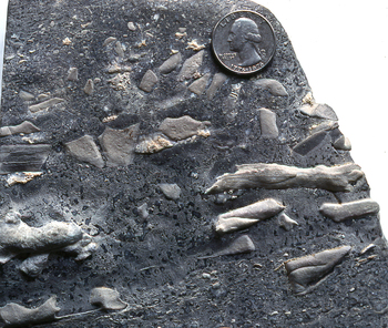
30.0 Skeletal limestone breccia. Saltville Quadrangle, Virginia. Skeletal fragments to whole fossils, as well as limestone clasts, make up the clast composition of this limestone–matrix breccia. Some smaller clasts, especially in the upper part of the sample, show significant rounding. [A 25-cent piece of 23 mm provides scale.] |
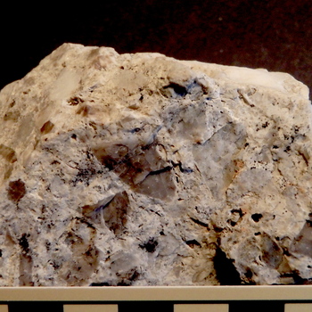
30.1 Chert breccia. Broadford Quadrangle, southwestern Virginia. Angular clasts of brownish-gray to white chert are the clasts in this white chert-matrix breccia. [Scale is standard GSA scale with cm divisions.] |
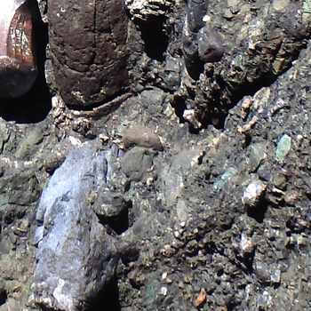
31.0 Polymict diamictite. Heaven's Beach, Sonoma Coast State Park, California. Clasts of various sizes (in outcrop, up to several meters in diameter) occur in a muddy, sandy matrix. Clasts visible here include blue glaucophane schist, green chert, green to grayish-green mafic metavolcanic rock, red-brown to gray sandstone, and white quartz. [Scale is provided by length of the half hammer-head in the upper left corner, which is about 6 cm.] |
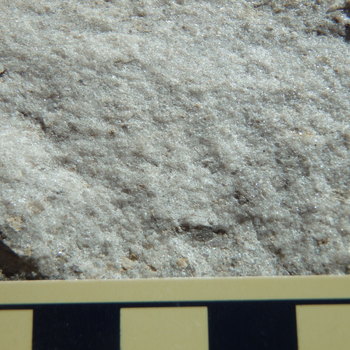
32.0 Quartz arenite. Tumbling Creek, Clinch Mountain Wildlife Management Area, Virginia. Moderately sorted, medium quartz sand grains, with a few granules of gray chert and buff-colored mudrock at the bottom of the field of view, constitute this rock. [Scale is standard GSA scale with cm divisions.] |
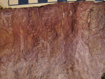
32.1 Hematitic quartz arenite. Clinch Mountain, Broadford Quadrangle, Virginia. Fine-grained quartz sand is cemented by red-brown to purple-maroon hematite in this rock. Some silica-cemented areas of quartz sand forming white lines in checker-like stacks represent Scolithus sp. fossil burrows. |
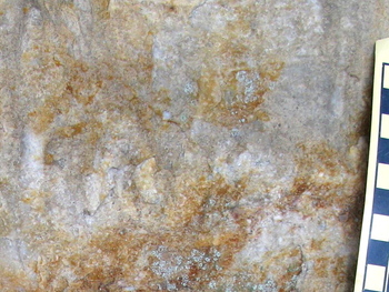
32.2 Granule-bearing quartz arenite. Clinch Mountain, Broadford Quadrangle, Virginia. Medium-coarse quartz sand in this view contains some granules of quartz (e.g., in the center of the photo) and is cemented locally by yellow-brown limonite and pink to red hematite. Greenish –gray patches are lichens growing on the rock. [Scale is standard GSA scale with cm divisions.] |
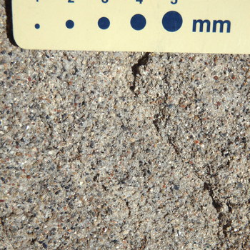
32.3 Lithic quartz arenite (sandstone). Dinosaur National Monument, Utah. Medium- to coarse-grained, moderately sorted sand of variously colored quartz, plus rock fragments of different types, including black, red, and other colors of chert, comprise this sandstone. Compare to granule conglomerate in 29.0, with which it is associated. [Scale is standard GSA scale with mm diameter dots.] |
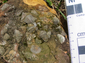
32.4 Fossiliferrous quartz arenite. Clinch Mountain north of Tannersville, Broadford Quadrangle, Virginia. Brachiopod fossils decorate the bedding surface of this limonite- and silica-cemented, fine-grained quartz arenite. |
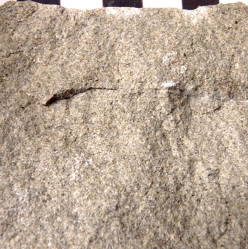
33.0 Feldspathic arenite. Lone Tree Creek Quadrangle, California. A few black biotite, chert, and lithic clasts, plus cream-colored feldspar grains and variously colored, but dominantly gray, glassy quartz grains, dominate this sandstone. A small amount of hematite cement gives the rock a light pink cast. [Scale is standard GSA scale with cm divisions.] |
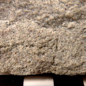
33.1 Feldspathic arenite. Near Holbrook, Arizona. Cream-colored feldspar—and variously colored, but dominantly gray glassy quartz—dominate this sandstone, which also contains black lithic clasts. Hematite cement gives the rock a pink cast. [Scale is standard GSA scale with cm divisions.] |
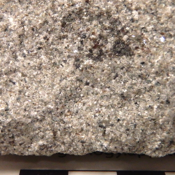
34.0 Lithic feldspathic wacke. West Virginia. Clay matrix binds dark gray to black lithic clasts (one of which is a large dark gray clast in the upper right center), white feldspar grains, and gray quartz grains in this sandstone. [Scale has cm divisions.] |
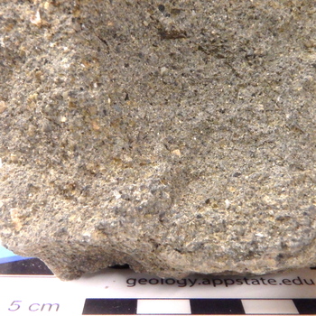
35.0 Granular feldspathic lithic wacke. Rutherford, California. White and yellow-brown feldspar grains, gray quartz, and brown to black lithic fragments occur in a gray matrix of clay minerals. |
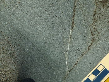
35.1 Granule-bearing, feldspathic lithic wacke. Sonoma Coast State Park, north of Jenner, California. Black lithic fragments of various sizes, including granule to pebble-size shale chips, occur as clasts, with gray to gray-green quartz and white feldspars, in a greenish-gray matrix of chlorite (?) and clays. [Scale has cm divisions.] |
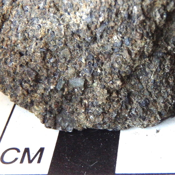
35.2 Granular lithic wacke. Heaven’s Beach, Sonoma Coast State Park, California. Abundant lithic fragments of shale, green chert, and other rock types occur with very coarse, sand-sized grains of brown, pink, and gray quartz and white feldspars in a mud matrix with local calcite cement. [Scale has cm divisions.] |
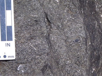
35.3 Pebble-bearing, granular lithic wacke. Blind Beach, Sonoma Coast State Park, California. Fluid-induced, soft sediment deformation modified the fabric of this pebble-bearing sandstone. Black mudrock fragments and brown to gray sandstone clasts comprise the pebbles. The sand grains consist of black lithic clasts, white feldspars, and gray quartz in a gray, mud matrix. Patches of deformed white calcite cement and deformed lenses and layers of dark gray mudrock are prominent in the field of view. |
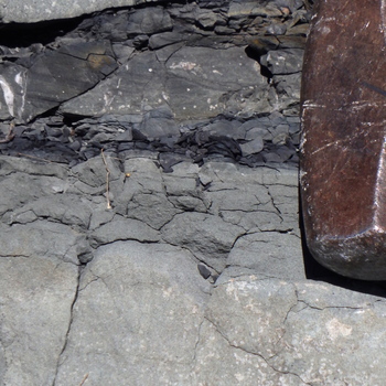
36.0 Siltstone (light gray) and mudshale (dark gray) occur between dark gray lithic wacke (above) and medium to light gray lithic wacke sandstone (below). Heaven's Beach, Sonoma Coast State Park, Sonoma County, California. [Scale is provided by 12 cm hammerhead in upper left.] |
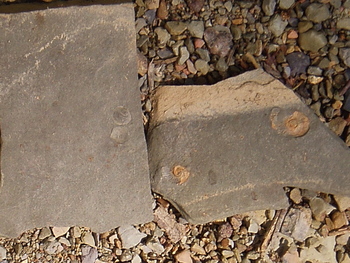
37.0 Mudrock with ammonoid cephalopods and brachiopods. Highway 91, Broadford Quadrangle, Virginia. |
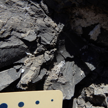
38.0 Siliceous shale with gypsum veins. Dinosaur National Monument, Utah. |
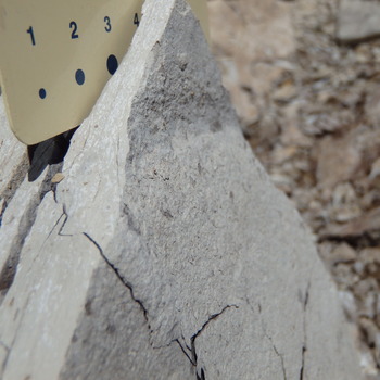
38.1 Shale. Duchesne, Utah. [The scale is the standard GSA scale with mm and inch divisions.] |

39.0 Lime mudstone. Locality near Ciegnes, France. [Scale has cm divisions.] |
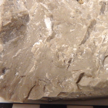
40.0 Skeletal wackestone. Guadalupe Mountains, Texas. Small white to clear calcite fossil fragments are scattered within a light brown lime mud matrix in this wackestone. [Scale has cm divisions.] |
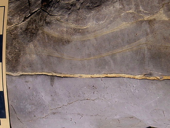
40.1 Wackestone (below) and lime mudstone (above). Ward Cove, north-central Broadford Quadrangle, Virginia. The wackestone at the bottom of the picture contains little fragments of fossils and limestone in a gray lime mud matrix. The uppermost layers, showing flame structures, are lime mudstone. [Scale is standard GSA scale with cm divisions.] |
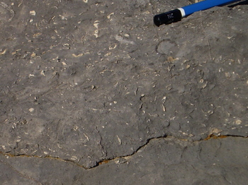
41.0 Packstone-Wackestone. Ward Cove, north-central Broadford Quadrangle, Virginia. Cream-colored and gray fossils and fossil fragments are immersed here in a dark gray lime mud matrix. Where very abundant and commonly touching, the abundant fossils render the rock a packstone. Locally, where the fragments are less abundant, the rock is a wackestone. [Scale is provided by the 1.8 cm black end of a pencil.] |
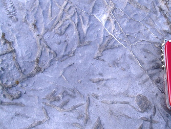
42.0 Fossiliferous packstone. Southeastern Broadstone Quadrangle, Virginia. Bryozoans lie on and below the bedding surface of this fossiliferous packstone with gray lime mud matrix. [Scale is provided by the approximately 7 cm long knife at right.] |
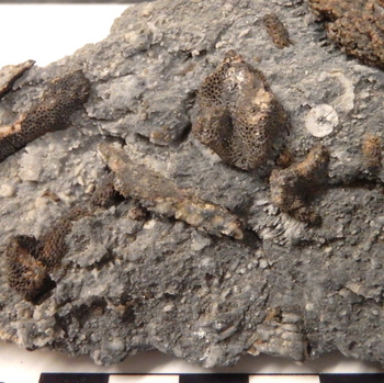
42.1 Skeletal Packstone. Vaughn Gulch, Inyo Mountains, California. Brown weathered bryozoans and white crinoid fragments are clasts in the recrystallized, dark gray matrix of this packstone. [Scale shows cm divisions.] |
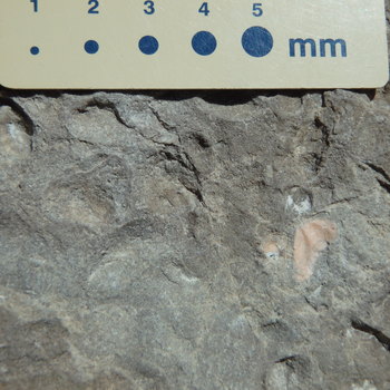
43.0 Sandy skeletal packstone with pelecypods. Dinosaur National Monument, Utah. The gray, sand-coated bumps and white to pink shells are pelycepod shells in this sandy limestone. |
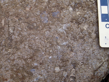
44.0 Fossiliferous grainstone. Southwestern Saltville Quadrangle, Virginia. A diverse group of fossils and fossil fragments are bound by limestone matrix in this weathered grainstone with hematitic coating. [Scale is standard GSA scale with cm divisions.] |
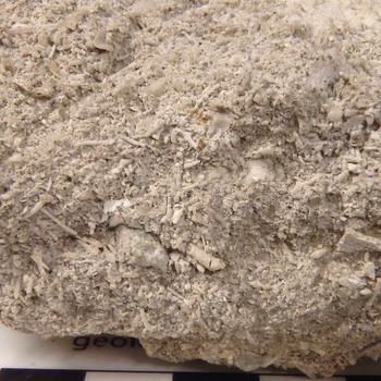
45.0 Skeletal Grainstone. Castle Hayne, North Carolina. A little calcite cement binds the fossil skeleton fragments in this grainstone. [Scale shows cm divisions.] |
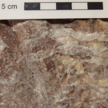
46.0 Hematitic bryozoan crystalline boundstone. Knoxville, Tennessee. Algal layers, recrystallized and partly stained by red hematite comprise this boundstone. |
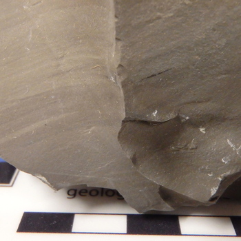
47.0 Petroliferous dolomudstone. Tucker, Utah. [Scale shows cm divisions.] |
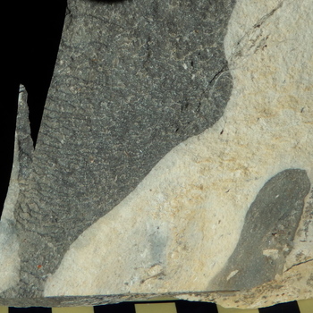
47.1 Dolostone (white) with gray chert. West of Junction City, Flint Hills, Kansas. [Scale shows cm divisions.] |
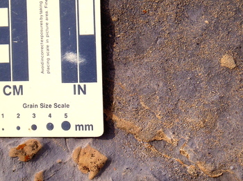
47.2 Dolomitic ooid packstone. Southern Broadstone Quadrangle, Virginia. Cream-colored and iron-oxide-stained, weathered ooids and small fossil fragments in a gray dolomitized lime mud matrix characterize this dolomitic packstone. Below the scale, where the dolomitized lime mud dominates, the rock is a dolowackestone. [Scale is standard GSA scale with cm divisions.] |
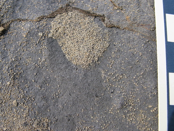
47.3 Ooid dolopackstone and dolowackestone. Southern Broadstone Quadrangle, Virginia. Cream-colored, weathered ooids and an ooid clump occur in dark gray dolomite. Area of sparse ooids is ooid dolowackestone. [Scale is standard GSA scale with cm divisions.] |
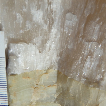
48.0 Calcite-aragonite travertine. Fairfield, California. Laminations of aphanitic crystalline limestone (below) with acicular, aragonite crystalline limestone (above). [Scale shows mm divisions.] |
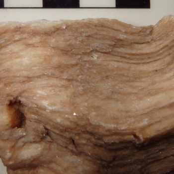
49.0 Gypsum evaporite. Fibrous. Saltville, Virginia. [Scale shows cm divisions.] |

50.0 Bituminous coal. Near Pineville, Kentucky. [Scale shows cm divisions.] |
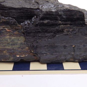
50.1 Bituminous coal. Morgantown, West Virginia. [Scale shows cm divisions.] |
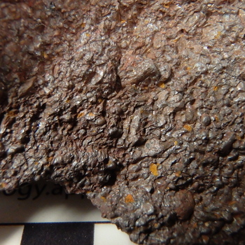
51.0 Skeletal oolitic ironstone. Birmingham, Alabama. [Scale shows cm divisions.] |
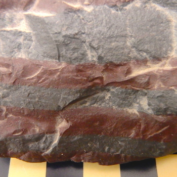
52.0 Banded iron formation. Minnesota. Gray-colored, aphanitic, iron-oxide-rich mineral layers interlayered with iron-rich red chert. [Scale shows cm divisions.] |
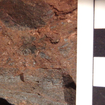
52.1 Banded iron formation breccia. Dickinson County, Michigan. Layers and fragments of gray magnetite-hematite-rich banded iron formation are cemented by hematite cement. [Scale shows cm divisions.] |
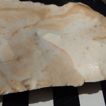
53.0 Opaline chert. Kenwood, California. [Scale shows cm divisions.] |
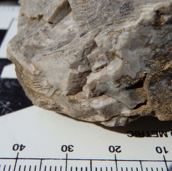
53.1 Chert. Broadford Quadrangle, Virginia. Laminated white chert. This white chert replaced dolostone, which replaced laminated limestone. Black and yellow dots are lichens growing on the surface of the rock. [Scale shows mm and cm divisions.] |
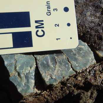
53.2 Green Chert. Marin County, California. This waxy to crystalline green chert is actually a clast in an olistostromal metawacke. [Scale is standard GSA scale with mm dots and cm divisions.] |
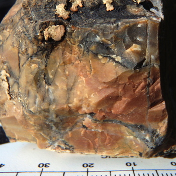
53.3 Yellow-red radiolarian chert. NE Diablo Range, California. Goethite (?) and hematite color the chert, which has hints of radiolaria appearing as gray spots in the yellow-red chert on the right edge. The black veins are manganese oxide mineral veins, originally deposited as manganese minerals with the chert and remobilized into veins. Gray quartz veins also cross the chert bed and the included, faint laminations. Cream-colored to reddish lichens are present on the upper surface. [Scale shows mm and cm divisions.] |
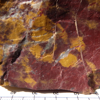
53.4 Yellow-red chert. Aliso Canyon, California. Yellow limonite and red hematite color this chert. [Scale shows mm and cm divisions.] |
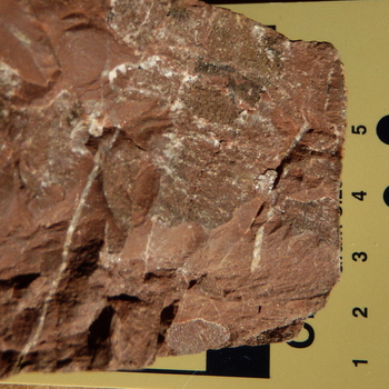
53.5 Red crystalline chert. NE Diablo Range, California. White veins of crystalline quartz cut across this grainy, recrystallized chert layer. Black areas are manganese oxide minerals. [Scale is standard GSA scale with cm divisions.] |
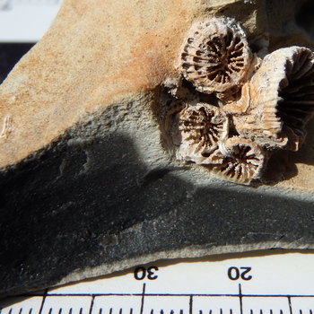
54.0 Black fossiliferous chert. England. A weathered surface colored by yellow orange limonite or goethite covers the coral-bearing black chert (typically called “flint”) of this sample. [Scale shows mm and cm divisions.] |
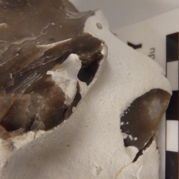
54.1 Black opaline chert with a white chalk coating. Seaford, Sussex, England. [Scale shows cm divisions.] |
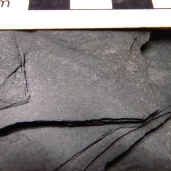
55.0 Dark gray slate. Central Virginia. This aphanitic rock displays typical slaty structure and a slight red coloration, probably due to oxidation of iron-bearing minerals to hematite. [Scale shows cm divisions.] |
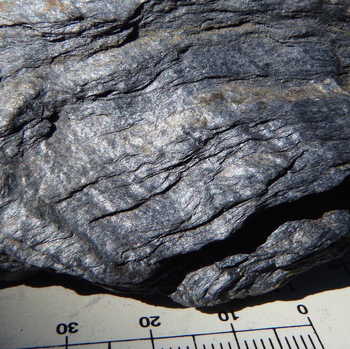
56.0 Glaucophane phyllite. Healdsburg Quadrangle, California. This sample displays classic phyllitic structure with wavy, aphanitic to very fine-grained layers of minerals, in this case, predominantly blue glaucophane and silvery-white white mica (muscovite-paragonite). A few tiny red garnets are present. [Scale shows mm and cm divisions.] |
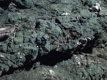
57.0 "Greenstone" (metabasalt). Pillowed. Rock Point, Sonoma Coast State Park, California. The rock is aphanitc and green, hence the name “greenstone.” In this picture, rounded pillows are cut locally by shear planes and white calcite veins. [Pillows measure 10 cm to 30 cm. The thin gray layer at left is about 2 cm thick, for scale.] |
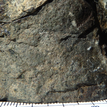
57.1 Metabasalt (“greenstone”). Nicasio Reservoir, Marin County, California. A few dark green augite and white plagioclase phenocrysts are the only visible grains in this fine-grained to aphanitic metavolcanic rock. [Scale shows mm and cm divisions.] |

58.0 Metachert with pumpellyite. California Coast Ranges. Green-colored metachert with aphanitic pumpellyite. Gray quartz veins cut across the chert. [Scale shows mm and cm divisions.] |
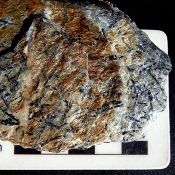
58.1 Metachert with ferro-glaucophane and white mica. Jenner, California. Limonite (yellow) stains the white mica that defines some foliation planes. The bluish-black needles on foliation planes are ferro-glaucophane. [Scale shows cm divisions.] |
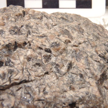
58.2 Stilpnomelane-garnet metachert. NE Diablo Range, California. Brownish-black sheets of stilpnomelane are visible, but microscopic garnets within the recrystallized quartz of the chert make the metachert pink in color. Some patches of gray quartz without garnet are visible. [Scale shows cm divisions.] |
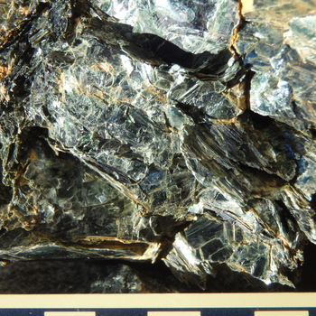
59.0 Chlorite diablastite. Frank, North Carolina. [Scale shows cm divisions.] |
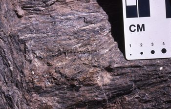
59.1 Anthophylite diablastite. Todd, North Carolina. [Scale is standard GSA scale with cm divisions.] |
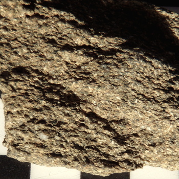
60.0 Semi-schistose jadeite-lawsonite metawacke. Hospital Creek area, NE Diablo Range, California. Some gray, glassy quartz grains, a few green chlorite grains, and many white grains that are plagioclase grains partly to entirely replaced by jadeite (pyroxene) are visible. Microscopic and X-ray analyses indicate that the goethite (?)-stained matrix material is a mix of glaucophane, chlorite, white mica, and chlorite-vermiculite minerals. [Scale shows cm divisions.] |
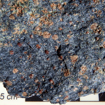
61.0 Garnet-glaucophane schist. Jenner, California. Omphacite (green) and garnet in the upper left represent a relict of eclogite that is being replaced by a garnet-glaucophane assemblage that is the dominant mineralogy of the replacing glaucophane schist. Some green minerals associated only with glaucophane and garnet (e.g., in the middle right) may be epidote. [Scale shows cm divisions.] |
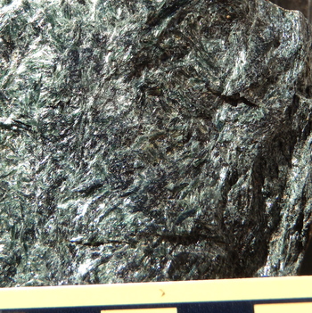
61.1 Chlorite-actinolite schist. NE Diablo Range, California. Chlorite (black appearing flaky minerals) and actinolite (green acicular minerals) comprise this schist. [Scale shows cm divisions.] |
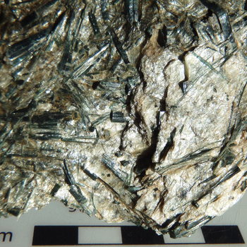
61.2 Actinolite-talc schist. NE Diablo Range, California. White talc encloses green, acicular actinolite crystals. [Scale shows cm divisions.] |
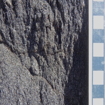
61.3.1 Quartz-biotite schist. Lake Alpine area, Highway 4, California. Quartz is white and biotite is black. Compare with photo of different orientation in 61.3.2 . [Scale is standard GSA scale with mm diameter dots.] |

61.3.2 Quartz-biotite schist. CA Highway 4 near Lake Alpine, California. Quartz is white and biotite is black. Compare with photo of different orientation in 61.3.1. [Scale shows cm divisions.] |
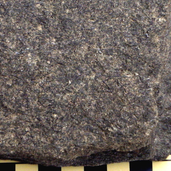
61.4 Hornblende-biotite schist. Michigan. Rock is composed of black needle-like grains of hornblende, brownish-black sheets of biotite, white feldspars, and gray quartz (only a few grains of quartz are visible). Sample courtesy of Sonoma State University Geology Department. [Scale shows cm divisions.] |
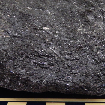
61.5 Hornblende schist. Mitchell County, North Carolina. Black acicular hornblende with minor white plagioclase, gray quartz, and green epidote (upper left) comprise the rock. [Scale shows cm divisions.] |
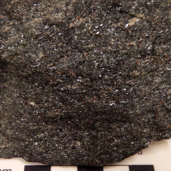
61.6 Garnet-hornblende schist ("amphibolite"). Ring Mountain, California. Small pink garnets, white plagioclase feldspar, and black, acicular hornblende constitute the bulk of this rock. A green omphacite vein cuts the rock in the upper left. [Scale shows cm divisions.] |
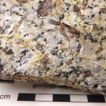
61.7 Garnet-staurolite-biotite-quartz-white mica schist. Highway 221, Ashe County, North Carolina. Red equant garnet, brown rectangular staurolite, black biotite, gray quartz, and silvery-white white mica are the major minerals of this rock. [Scale shows cm divisions.] |
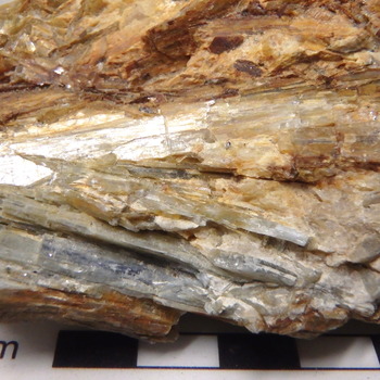
61.8 Quartz-kyanite schist. Brown’s Creek, Celo, North Carolina. Gray to white quartz and white to blue kyanite are the two major minerals of this rock, but white alkali feldspar, silvery white mica, tiny metallic black ilmenite (?), and red-brown iron oxides are minor constituents. [Scale shows cm divisions.] |
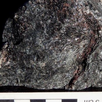
61.9 Alkali-feldspar-bearing, garnet-quartz-biotite-sillimanite schist. Winding Stair Gap, North Carolina. Minor pinkish-white alkali-feldspar forms lenses within this schist of red garnet, gray quartz, black biotite, and silvery-white to gray needle-like sillimanite. |
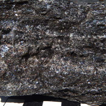
62.0 Riebeckite-quartz-garnet-stilpnomelane gneiss. Laytonville, California. Light colored bands are gray quartz or gray quartz and pink garnet, whereas dark colored bands are dominated by stilpnomelane with garnet. [Scale shows cm divisions.] |

62.1 Quartz-plagioclase-glaucophane gneiss. Austin Creek, California. Bluish-black glaucophane dominates dark bands, whereas white plagioclase and gray quartz dominate the light-colored bands. [Scale shows cm divisions.] |
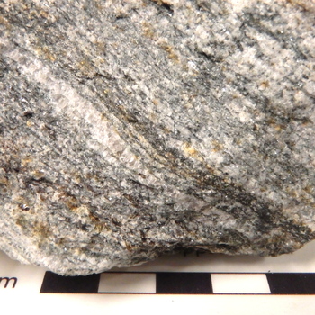
63.0 Biotite-gedrite-quartz-plagioclase gneiss. Booneford, North Carolina. Black biotite and greenish gray gedrite form dark areas, whereas white plagioclase and gray quartz dominate the light areas of this rock. Yellow limonite stains some grains. [Scale shows cm divisions.] |
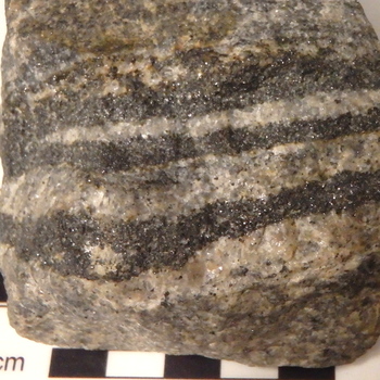
63.1 Magnetite-quartz-feldspar gneiss. Llano County, Texas. Dark bands are magnetite-rich, whereas light bands are alkali feldspar- (white) and quartz- (gray) rich. Yellow limonite stains some grain surfaces. [Scale shows cm divisions.] |
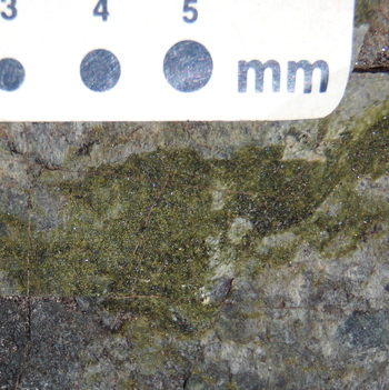
64.0 Epidote hornfels. Locality is east of Alpine Lake, California along CA Highway 4. Granular yellow-green to dark green epidote is associated here, at a granite-biotite schist contact, with gray quartz, plus associated tiny black grains of biotite, and white alkali feldspar. Secondary, red hematite provides some red-colored coatings. Thin veins of unknown composition cut the sample. |

65.0 Bedded hematitic aragonite marble. NE Diablo Range, California. The carbonate grains in this marble are aragonite, which appears to be converting to calcite. Cleavages are especially visible in the upper right, but the grain size in the lower part of the sample is clearly aphanitic. Hematite provides the red color. White to gray calcite veins cut across the sample, composed of an upper and a lower bed, separated by a chlorite coated (greenish-gray) bedding plane. |
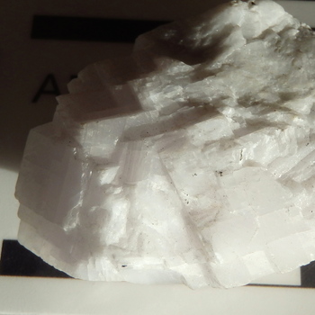
65.1 Calcite marble (coarse-grained). Santa Cruz, California. The rock is composed of white calcite, within the grains of which there are tiny, unidentified dark green minerals and reflective black grains, probably of graphite. [For scale, the field of view is about 4.25 cm wide.] |
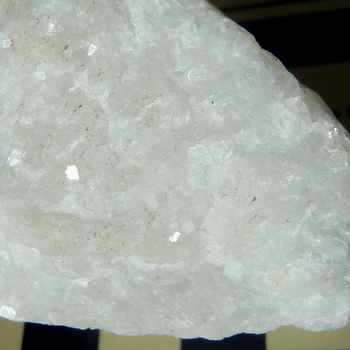
65.2 Dolomite marble. Locality unknown. White dolomite constitutes the entire rock, except for a few tiny patches of brown clay on the surface. [Scale shows cm divisions.] |

65.3 Rhodochrosite marble. Buckeye Mines, NE Diablo Range, California. Pink rhodochrosite constitutes most of this rock, but veins and coatings of black manganese oxide minerals and some yellow-orange limonite coatings complete the mineralogy of the specimen. [Scale shows cm divisions.] |
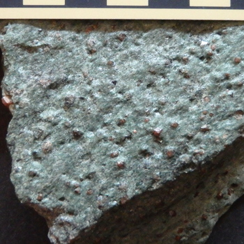
66.0 Eclogite. Reed Station, Tiburon Peninsula, California. Orange and red-to- black-appearing garnets occur with granular omphacites (clinopyroxenes), constituting this rock. A few small, blue, acicular grains of glaucophane are present locally. [Scale shows cm divisions.] |
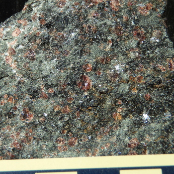
66.1 Hornblende eclogite. Bakersville, North Carolina. Dark green to black hornblende replaces medium green omphacite. Red-brown garnets occur with both minerals and in the upper right area accompanied by white plagioclase. Yellow-brown limonite replaces some omphacite and garnet. [Scale shows cm divisions.] |
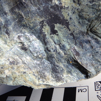
67.0 Serpentinite. (Blue-green). Locality near Cloverdale, California. Blue-green serpentinite with minute black magnetite grains and coatings of yellow limonite and black manganese oxide minerals. Surfaces show slickensides. |
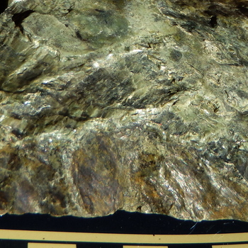
67.1 Serpentinite. Locality near Cloverdale, California. Yellow-green to green serpentinite shows some cross fiber fabric and slickensides. Yellow-orange iron oxides/hydroxides (limonite or goethite) coat the lower surface. Black magnetite is abundant. [Scale shows cm divisions.] |
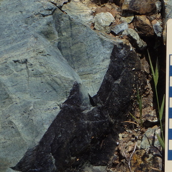
67.2 Serpentinite gneiss. Bolinas-Fairfax Road, near Azalea Hill, Marin County, California. Aligned light yellow-green to darker blue-green serpentinite form a gneiss with black serpentinite veins oriented generally parallel to the foliation. Additional veins occur in an orientation generally perpendicular to the foliation. Analyses of similar blocks of serpentinite showed the serpentine to be antigorite. [Scale is standard GSA scale with cm divisions.] |
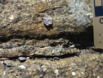
68.0 Serpentinized harzburgite. Del Puerto Canyon, California. Serpentine minerals show up as green to black aphanitic materials and locally appear white, where light is reflected from them. The dominant fabric is one of a cross-fracture pattern, with dark serpentine veins filling the fractures that surround little, less serpentinized, cm-scale blocks. Some square grains distinctly visible because of reflection from relict cleavage (e.g., lower left and lower right), are serpentinized orthopyroxenes usually referred to as bastites. [Scale is standard GSA scale with cm divisions and mm-scale dots.] |
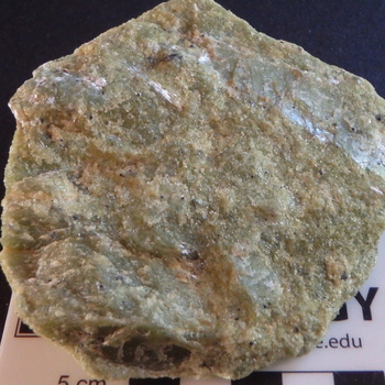
68.1 Metadunite. Day Book Mine, western North Carolina. Large, deformed porphyroclasts of olivine with flat parting surfaces are surrounded by a matrix of fine-grained, recrystallized olivine with scattered grains of chromium spinel (note the tiny chromite grain in the center with the perfectly triangular face of an octahedron), talc (white to silvery-white), and tremolite (e.g., upper center, white to light green). Note the modest fabric produced by an upper left to lower right grain orientation. |
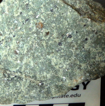
68.2 Metaharzburgite with tremolite vein. Day Book Mine, North Carolina. Fine-grained olivine (green) dominates the rock, which contains brown to black orthopyroxenes, silvery-white talc, silvery-white chromium chlorite (kammererite) associated with black chromium spinels, and a vein of brownish-green tremolite (lower left). Notice the faint grain elongations in an upper right to lower left orientation. [Scale shows cm divisions.] |

68.3 Metaorthopyroxenite. Rich Mountain, near Boone, North Carolina. Stretched and bent grains of brown orthopyroxene dominate this rock. A few grains of olivine (green) are present (e.g., near right middle edge) and a patch of yellow limonite is present in the lower right quadrant. [Scale shows cm divisions.] |
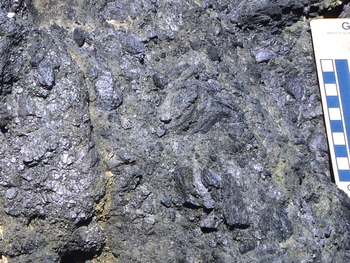
69.0 Fault breccia composed of glaucophane schist. Heaven’s Beach, Sonoma Coast State Park, California. Blue to black, angular fragments of glaucophane schist occur in a matrix of blue-gray gouge. [Scale is standard GSA scale with cm divisions.] |
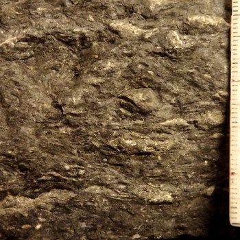
70.0 Quasimylonite. NE Diablo Range. Lenses down to micro-lenses (microlithons) of sandstone (light gray) “float” in a matrix of dark gray-brown, sheared mudrock. A few white quartz veins are also present. Shear fractures and slickensides (not visible) on shale surfaces suggest brittle failure and dislocation. “Necking” and elongation of sandstone lenses (e.g., at bottom of image) indicate plastic deformation of the sandstone layers. [Scale on right shows mm divisions.] |
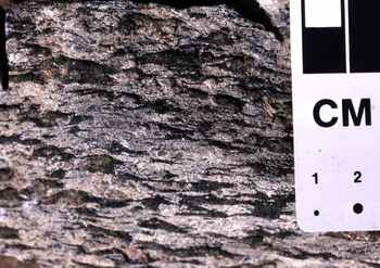
71.0 Hornblende metagabbro protomylonite. Todd, North Carolina. Elongate and highly deformed black hornblende grains are surrounded in this rock by abundant, highly deformed, white to gray plagioclase feldspars. [Scale is standard GSA scale with cm divisions and mm-scale dots.] |
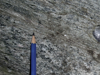
71.1 Mylonite zone in metaquartzarenite. Grandfather Mountain, North Carolina. Zone in upper right is mylonite derived from metamorphosed quartz arenite in lower left. Dark, microscopic, recrystallized grains of quartz and feldspar mixed in with chlorite and white mica in the mylonite surround syntectonically recrystallized lenses and bands of white to gray quartz and light pinkish-gray alkali feldspar. On the far right, a glassy-gray fragment of a quartz vein is surrounded by mylonitic materials. On the lower left, gray to white grains of quartz and pinkish-gray alkali feldspar with a recrystallized matrix of silvery-white, white mica and green to black chlorite, plus a few iron oxide grains, constitute the meta-sandstone. [Scale is provided by the approximately 1.5 cm sharpened end of the pencil.] |